MAKE AN APPOINTMENT TODAY!
Hand Anatomy
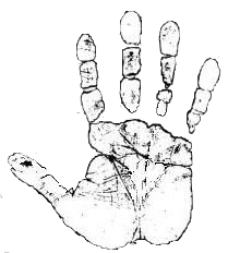
Hand anatomy and function are highly complex. The thumb alone is controlled by nine individual muscles innervated by the radial nerve, median nerve, and ulnar nerve. The thumb movement is so complex that there are six unique descriptive terms for describing only the direction of the thumb joint at the base of the thumb. If you play an instrument, such as the guitar or the piano, you are likely aware of how the slightest ache or pain affects your finger positioning required to play those instruments.
The hand structures belong to several categories:
The hand structures belong to several categories:
Bones and Joints
The hand and wrist have twenty-seven bones. Five metacarpal bones form the palm and connect to each finger and thumb. Each finger is formed by proximal phalanx, middle phalanx, and distal phalanx. The thumb has only a proximal phalanx and a distal phalanx.
The knuckle joint consists of metacarpophalangeal joints (MCP joints) and allows us to straighten all fingers and thumb simultaneously.
The proximal phalanges, middle phalanges, and distal phalanges in each finger are joined by an interphalangeal joint (IP joint). The interphalangeal joint closest to the MCP joint is the proximal IP joint (PIP joint). The distal IP joint (DIP joint) is the joint farthest away from the MCP joint. The thumb has one interphalangeal joint. IP joints allow us to bend and straighten fingers and thumb individually.
The wrist contains eight carpal bones. The wrist joint is formed by joining the carpal bones in the wrist with two forearm bones, the radius and the ulna.
All joints of the hand and wrist have shock-absorbing smooth-surface articular cartilage, which facilitates hand and wrist motion.
Ligaments and Tendons
Ligaments are fibrous connective tissue, which connects two bones. Collateral ligaments link two bones on either side of a joint and prevent unnatural lateral bending of each joint.
The volar plate ligament connects the proximal phalanx to the middle phalanx on the palmar side of the hand. It keeps the PIP joint from hyperextension.
The tendons connect muscles to bones. The extensor tendon arises from the forearm muscles and widens over the back of a phalanx to form an extensor hood, which connects to the middle and distal phalanges. The extensor tendons allow fingers to straighten. Central slip is the site of extensor tendon attachment to the middle phalanx.
Muscles
1. Carpal muscles act at the wrist and, in some cases, the elbow.
2. Extrinsic hand muscles are the long flexors and extensors and act on the wrist and the digits to control crude movements and produce a forceful grip.
3. Extrinsic thumb muscles act on the first digit.
4. Intrinsic hand muscles are responsible for the fine motor hand functions and act only on the digits.
5. Lumbricals are four muscles, each associated with a finger. They are crucial to finger movement, linking the extensor tendons to the flexor tendons.
6. Dorsal and palmar interossei muscles are located between the metacarpals. They abduct (dorsal interossei) and adduct (palmar interossei) fingers and assist the lumbricals in flexion at the MCP joints and extension at the IP joints.
7. Flexor retinaculum is a strong band of the fibrous ligament (material that connects bone to bone) stretches across the back of the wrist. It affects muscles that help flex the hand.
8. Extensor retinaculum ligament affects muscles that help extend fingers.
9. Pronator teres originates at the top of the humerus, crosses the forearm, and connects to the ulna. It helps turn the palm downward.
- Extensor Carpi Radialis Longus
- Extensor Carpi Radialis Brevis
- Extensor Carpi Radialis Longus
- Flexor Carpi Radialis
- Flexor Carpi Radialis
- Palmaris Longus
2. Extrinsic hand muscles are the long flexors and extensors and act on the wrist and the digits to control crude movements and produce a forceful grip.
- Extensor Digitorum
- Extensor Indicis (Proprius)
- Extensor Digit Minimi (Proprius)
- Flexor Digitorum Superficialis
- Flexor Digitorum Profundis
3. Extrinsic thumb muscles act on the first digit.
- Abductor Pollicis Longus
- Extensor Pollicis Brevis
- Flexor Pollicis Longus
- Extensor Pollicis Longus
4. Intrinsic hand muscles are responsible for the fine motor hand functions and act only on the digits.
- Thenar Muscles Opponens Pollicis, Abductor Pollicis Brevis, and Flexor Pollicis Brevis produce a bulge at the base of the thumb known as the thenar eminence.
- Hypothenar Muscles Opponens Digiti Minimi, Abductor Digiti Minimi, and Flexor Digiti Minimi Brevis produce the hypothenar eminence at the base of the little finger.
5. Lumbricals are four muscles, each associated with a finger. They are crucial to finger movement, linking the extensor tendons to the flexor tendons.
6. Dorsal and palmar interossei muscles are located between the metacarpals. They abduct (dorsal interossei) and adduct (palmar interossei) fingers and assist the lumbricals in flexion at the MCP joints and extension at the IP joints.
7. Flexor retinaculum is a strong band of the fibrous ligament (material that connects bone to bone) stretches across the back of the wrist. It affects muscles that help flex the hand.
8. Extensor retinaculum ligament affects muscles that help extend fingers.
9. Pronator teres originates at the top of the humerus, crosses the forearm, and connects to the ulna. It helps turn the palm downward.
Nerves
The hand muscles are innervated by the radial, median, and ulnar nerves from the brachial plexus.
The radial nerve allows us to straighten and raise elbows, wrists, hands, and fingers. It provides touch, pain, and temperature sensations to portions of the back of the upper arm, forearm, and the back of the hand and fingers.
The median nerve innervates forearm muscles and facilitates bending and straightening of wrists, thumbs, and first three fingers. It allows forearm rotation and the downward turning of the palm. The median nerve is responsible for touch, pain, and temperature sensations to the:
- Palm side of the thumb, index and middle fingers, and part of the ring finger
- Forearm
- Thumb side of the palm
- Dorsal side of the index and middle fingers
The ulnar nerve controls nearly all of the small muscles in hand. It allows for (1) bending and straightening of the small and ring fingers, (2) gripping and holding items, and (3) performing fine motor tasks like writing with a pen, buttoning a shirt. As a sensory nerve, the ulnar nerve gives feeling to the small finger, side of the ring finger closest to the small finger, palm, and back of the hand on the small finger side.
Blood Vessels
The radial and ulnar arteries and their branches provide hand and wrist blood supply.
Disclaimer and Privacy
IZADIHAND.COM © 2011-2022 Kayvon David Izadi MD - All Rights Reserved
Webmaster
IZADIHAND.COM © 2011-2022 Kayvon David Izadi MD - All Rights Reserved
Webmaster
